Understanding an atomized flame requires more than observing its overall shape or brightness. Researchers may need to compare radiation signatures at multiple locations within the flame and relate those patterns to the reactions occurring there. Hyperspectral imaging for flame analysis brings spatial and spectral information together in one dataset, making it useful for combustion studies involving boron-based nanofluid fuels.
Why spatial spectral data matters in combustion research
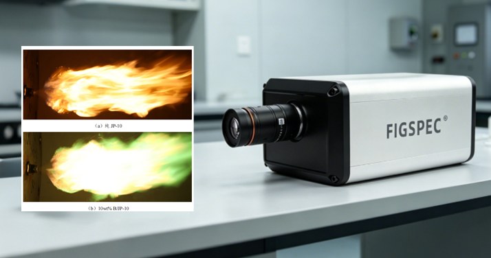
Boron-based high-energy nanofluid fuels have attracted interest in aerospace propulsion research because their atomization and combustion characteristics require detailed investigation. In a study of B/JP-10 nanofluid fuel, the objective was to measure the spatial characteristic emission spectra of the atomized combustion flame.
Conventional spectral measurement approaches can be limited when the goal is to compare spectral information from different parts of a flame. An imaging hyperspectral camera, by contrast, records the target's spatial information together with its spectral information. This enables analysis of how radiation intensity and characteristic spectral features change across the flame field.
The study used the FS-22 imaging hyperspectral camera from CHN SPEC with a nanofluid fuel atomization combustion test system. The system included sample feeding, an atomization nozzle, testing equipment, and sampling equipment. An air atomization nozzle atomized the fuel, while a plasma arc ignited the resulting sample jet.
Characteristic bands selected for B/JP-10 flame analysis
The camera collected spatial radiation spectral data from the atomized flame. Two radiation bands were selected according to characteristic spectra associated with boron and hydrocarbon fuel combustion:
- 431 nm: This blue band corresponds to CH radical radiation and was used to characterize the combustion reaction of the hydrocarbon fuel JP-10.
- 581 nm: This green band corresponds to BO₂ radical radiation and was used to characterize the combustion reaction of boron particles.
Examining the intensity distribution at these two wavelengths helps distinguish the predominant reactions in different flame locations. Rather than treating the flame as a single uniform region, researchers can assess localized patterns along its centerline and around its outer edges.
What the spectral observations indicated
Changes along the axial centerline
At the axial center of the atomized torch, the recorded spectra showed distinct changes by position. The spectral curves at Position 1 and Position 2 included the characteristic “five-finger peaks” associated with boron combustion. Radiation intensity increased with distance from the nozzle in this portion of the flame.
These observations indicated that boron combustion was present from the nozzle through Position 2 and became stronger as the boron particles moved downstream. From Position 3 through Position 5, the boron characteristic peaks were no longer observed at the flame center, indicating no significant boron-particle chemical reaction in that central section.
Differences across radial positions
A separate comparison examined radial positions around Position 4, where centerline radiation intensity was highest. Boron-related radiation peaks appeared at both the upper and lower edges of the atomized torch. However, total radiation intensity was slightly higher at the upper edge.
The study associated this difference with upward movement of JP-10 vapor under buoyancy, which increased the amount of JP-10 participating in reactions in the upper flame region. Boron-related peaks also remained evident at the lower edge, consistent with the downward movement of boron under gravity. Together, these results show how hyperspectral imaging can reveal non-uniform reaction behavior that may not be apparent in a single integrated measurement.
Combustion-zone interpretation
Using the spatial spectral radiation data together with atomization combustion images, the research team divided the center of the B/JP-10 atomization flame along the nozzle axis into four zones:
- B/JP-10 coupled combustion zone at the outlet section
- JP-10 single-phase combustion zone in the stable combustion section
- B/JP-10 coupled combustion zone in the tail flame section
- Boron single-phase combustion zone
This zone-based interpretation provides a structured way to investigate the atomization and combustion mechanism of the fuel. It also illustrates the value of combining image location with radiation spectra when comparing flame regions.
FigSpec FS-22 for combustion imaging studies
The FigSpec FS-22 imaging hyperspectral camera was used to collect the spatial radiation spectra described in this case. Its stated specifications are a 1920*1920 image resolution, a spectral range of 400-1000nm, a spectral resolution of 5nm FWHM, and 600 spectral channels.
For researchers studying atomized fuel flames, this type of measurement approach can support evaluation of component-related radiation distributions and combustion-zone behavior. In the B/JP-10 case, the combined spatial and spectral dataset supported analysis of CH and BO₂-related radiation bands across the flame, contributing to combustion mechanism research, fuel formulation studies, and combustion model development.