Early identification of rice bacterial leaf blight is important because visible symptoms may not be apparent during the initial stage of infection. Conventional visual field inspection can therefore be limited when leaves are still asymptomatic. Hyperspectral imaging for rice disease detection offers a different approach: it combines spatial imagery with spectral information to examine subtle physiological and biochemical changes in plant leaves.
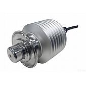
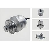
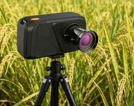
In one research application, the FigSpec FS-IQ-VISNIR portable hyperspectral camera was used to collect spectral data from rice leaves at healthy, mildly infected, and severely infected stages. The work illustrates how non-contact spectral imaging can provide data for feature extraction and intelligent classification workflows in crop research.
Why early spectral detection matters
Rice bacterial leaf blight can affect yield and food security. Once clear lesions are present, opportunities for timely prevention and control may be reduced. In contrast, hyperspectral imaging can capture information beyond what is readily observed through standard visual assessment.
This capability is relevant to agricultural research and to food-related supply chains that depend on plant health monitoring. Rather than relying on a single image channel, a hyperspectral system records spectral responses across many narrow bands. These responses can be processed to investigate leaf conditions and identify patterns associated with infection severity.
Data collection with FigSpec FS-IQ-VISNIR
The study used the FigSpec FS-IQ-VISNIR hyperspectral camera, which covers a spectral range of 400–1000 nm and has a spectral resolution of 2.5 nm. The stated image resolution is 1920 × 1920, with 1200 spectral channels.
Data were collected outdoors on sunny days between 10:00 and 14:00. The lens was positioned approximately 60–80 cm from the rice canopy. Exposure time was adjusted in real time to keep DN values in the 3000–4000 range, with the aim of limiting the influence of overexposure and noise.
The samples were divided into three conditions: healthy leaves, mildly infected leaves in an asymptomatic stage, and severely infected leaves. This comparison enabled the research team to examine spectral differences across disease levels. Because the camera supports fast, non-contact imaging, the approach can be used for both controlled collection and in-situ field-oriented data gathering.
Preprocessing and selection of informative bands
Raw hyperspectral data require processing before model development. In this application, the workflow included dark current correction, white reference correction, and Savitzky-Golay smoothing. Low signal-to-noise bands at the two spectral ends were removed, leaving 243 bands for modeling analysis.
Deep learning methods were then used to identify spectral bands that were more sensitive to bacterial leaf blight. The selected regions were concentrated around the green peak at 520–550 nm and the red-edge region at 680–720 nm. The green peak was associated with changes in chlorophyll content, while the red edge was described as reflecting leaf cell structure and stress conditions.
The results indicate that a smaller set of core bands may preserve much of the useful discriminatory information. In the study, roughly 8% of the core bands were used to reduce data dimensionality while supporting model efficiency and recognition stability.
Recognition results and practical interpretation
Using selected core bands as model input, the reported classification accuracy exceeded 96% in this research task and performed better than direct use of the full spectrum. For unbalanced sample scenarios, generative expansion of minority samples was reported to improve overall model performance by 6%–13%.
These findings should be understood within the conditions of the specific study. They demonstrate the value of combining suitable acquisition practices, spectral preprocessing, band selection, and classification methods. They do not mean that one result automatically applies to every rice variety, location, growth stage, or field environment.
A data foundation for crop research
For researchers investigating non-destructive crop analysis, the FigSpec FS-IQ-VISNIR provides visible-to-near-infrared spectral coverage for examining weak leaf-level differences. Its portable format supports laboratory and field data collection, while the resulting spectral data can be incorporated into machine learning or deep learning workflows.
In this application, hyperspectral imaging for rice disease detection created a technical path from leaf acquisition to targeted feature mining and disease classification. The key lesson is not only the camera specification, but also the disciplined workflow: collect consistent data, correct and screen the spectra, focus on informative regions, and validate recognition models against defined sample groups.