Phantoms
医用画像の評価や医療機器の検証では、毎回同じ条件で比較できる再現性の高い基準体が欠かせません。そうした場面で重要な役割を果たすのがPhantomsです。臨床現場そのものを置き換えるものではありませんが、CT、超音波、MRIなどの装置特性を安定した条件で確認し、研究・品質管理・教育の各プロセスを支える実用的なツールとして広く用いられています。
このカテゴリでは、単純な試験ブロックではなく、軟部組織、骨、臓器、血流評価、解剖学的構造など、目的に応じて設計されたモデルが中心になります。選定では、形状だけでなく、対応モダリティ、模擬される組織特性、評価したい項目との適合性を見ることが重要です。
Phantomsが医用画像評価で重視される理由
ファントムは、生体組織や解剖構造の一部特性を模擬するために設計された試験体です。被写体のばらつきに左右されにくいため、装置比較、撮像条件の検討、品質確認、手技トレーニングなどで安定した評価を行いやすくなります。
B2Bの調達や運用の観点では、単に「撮れる」ことよりも、同一条件で繰り返し検証できることに価値があります。より広い検証環境の中では、患者シミュレーションとあわせて活用することで、装置評価の流れをより体系的に構成しやすくなります。
このカテゴリで見られる代表的なファントムの方向性
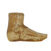
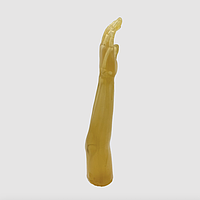
Phantomsの構成は多様で、用途によって求められる形状や再現レベルが異なります。たとえば新生児の頭部・腕・胴体のように部位に着目したモデルもあれば、ラットや犬を対象にした前臨床寄りのモデル、あるいはドプラ評価を意識した機能志向のモデルもあります。
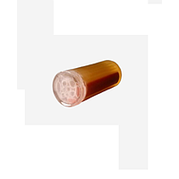
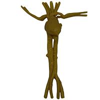
True Phantom Solutionsの製品群を見ると、その幅がわかりやすく表れています。True Phantom Solutions DP-C01 Doppler Phantom For X-Ray CT, US, MRI は流れや血管系を意識した評価に適し、True Phantom Solutions RT-C01 Rat Phantom (Cylindrical) For X-Ray CT, MRI は単純化された幾何形状で比較しやすい一方、True Phantom Solutions RT-A02 Rat Phantom (Anatomical) For X-Ray CT, US はより解剖学的な表現を重視した構成です。
モダリティ対応の違いが選定を左右する
実際の選定で最初に確認したいのが、対象となる画像モダリティです。このカテゴリには、X-Ray CTとUSに対応するもの、CTとMRI向けのもの、あるいはX-Ray CT、US、MRIの複数モダリティで使えるものが含まれています。同じ「ファントム」でも、評価したい信号やコントラストの種類が異なれば、適したモデルも変わります。
たとえば、新生児胴体モデルでも、True Phantom Solutions AN-N01 Newborn Torso (Advanced) For X-Ray CT, US と True Phantom Solutions AN-N02 Newborn Torso (Advanced) For X-Ray CT, MRI では想定される比較対象が異なります。超音波観察を含む運用か、CTとMRIの横断的な検証かによって、選ぶべき方向性は変わるため、導入前には運用目的を明確にしておくことが大切です。
形状だけでなく、模擬される材料特性にも注目
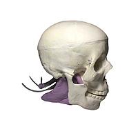
ファントム選定では、見た目の形状だけでは十分ではありません。骨、脳実質、軟部組織、皮膚、軟骨、臓器など、何を模擬しているのか、その材料特性が評価目的にどの程度関係するのかを確認する必要があります。密度、減衰、音速、硬さ、speckleの有無といった要素は、画像の見え方や比較結果に影響しやすいポイントです。
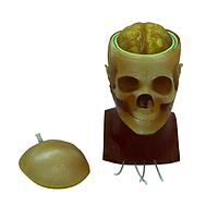
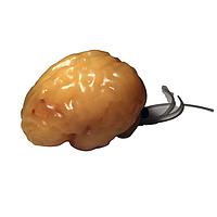
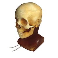
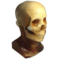
たとえば、True Phantom Solutions HD-N01 Newborn Head (Complex) For X-Ray CT, US, MRI、True Phantom Solutions HD-N02 Newborn Head (Simple) For X-Ray CT, US, MRI、True Phantom Solutions HD-N03 Newborn Head (Dynamic) For X-Ray CT, US, MRI は、いずれも新生児頭部をテーマにしながら、構成や評価の意図が異なるモデルです。また、True Phantom Solutions AM-S01 Newborn Arm For X-Ray CT, US, MRI は皮質骨と軟部組織を意識した比較的シンプルな部位モデルとして位置づけやすく、用途を絞った評価に向いています。
シンプルモデルと解剖学モデルの使い分け
シンプルな形状のファントムは、装置間比較や条件変更時の基礎確認に向いています。構造が整理されているため、結果の変化を読み取りやすく、研究初期や反復試験でも扱いやすいのが利点です。RT-C01のような円筒形モデルは、幾何学条件を明確にしながら評価したい場面で考えやすい選択肢です。
一方で、解剖学的な配置を持つモデルは、臓器位置関係や構造認識を含めた評価に向いています。RT-A02のようなアナトミカルモデルや、True Phantom Solutions DG-A02 Dog Phantom For X-Ray CT, MRI のようなより大きな動物モデルは、現実の撮像シナリオに近い条件を意識した検証で有用です。トレーニング、研究、プロトコル比較など、実運用に寄せた目的ではこちらが適する場合があります。
研究、品質管理、教育での活用場面
Phantomsは、研究開発、受入確認、定期的な品質保証、保守後の再確認、教育用途まで幅広く使われます。生体差の影響を抑えながら評価できるため、条件検討や装置の再現性確認に向いており、特に比較検証が多い現場で導入価値が高まります。
教育用途では、繰り返し観察やスキャンが必要な場面で有効です。新生児頭部や胴体モデルは小児・新生児領域を意識したトレーニングに適し、ラットや犬のモデルは前臨床研究や獣医系の検討にもなじみます。医療機器の総合的な点検体制を構築する場合は、輸液装置アナライザーや電気外科用アナライザーのような関連カテゴリも、運用全体の中で併せて検討されることがあります。
導入前に確認したい実務上のポイント
選定時には、まず「どのモダリティで使うのか」「どの部位または組織を模擬したいのか」「研究・QA・教育のどれが主目的か」を整理すると判断しやすくなります。そのうえで、解剖学的なリアリティを重視するのか、比較しやすい単純構造を優先するのかを見極めると、候補を絞り込みやすくなります。
また、サイズ感や取り回しも見落とせません。新生児向けの頭部・腕・胴体モデルのようにコンパクトなものは日常的な評価に組み込みやすく、犬モデルのような大型モデルは、より広い構造を含めた検証に向きます。製品名だけで判断するのではなく、使用シナリオとの整合で選ぶことが、導入後のミスマッチを減らす近道です。
運用に合ったPhantomsを選ぶために
このカテゴリのPhantomsは、汎用的な試験体を一括りにしたものではなく、評価対象や運用目的に応じて選ぶべき基準体の集合です。新生児頭部、腕、胴体、ラット、犬、Doppler用途など、対象が異なれば、求められる再現性や情報も変わります。
モダリティ対応、模擬材料の特性、構造のリアリティ、そして運用目的の4点を軸に見ていけば、必要な検証に適した一台を見つけやすくなります。Phantomsを適切に選定することは、画像評価の質を高めるだけでなく、日常の品質管理や研究の信頼性を安定させるうえでも重要です。
数量割引、まとめ買い価格の更新、新製品情報をメールでお届けします。
登録することで、当社の利用規約およびプライバシーポリシーに同意したものとみなされます。
認定専門家へ直接アクセス