Thiết bị hình ảnh điện tử y tế
의료 영상 시스템을 검토할 때 구매 담당자와 설계 엔지니어가 실제로 궁금해하는 것은 단순한 모델 목록이 아닙니다. 어떤 검출기 형식이 필요한지, 정지 영상과 동영상 촬영 중 어디에 초점을 둘지, 해상도와 전송 인터페이스가 시스템 통합에 어떤 영향을 주는지까지 함께 판단해야 합니다.
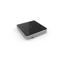
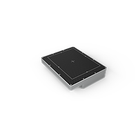
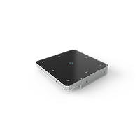
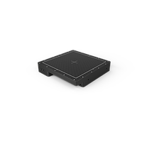
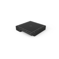
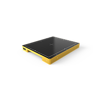
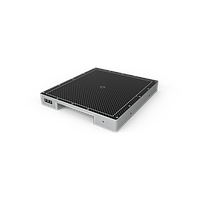
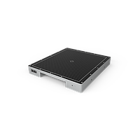
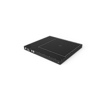
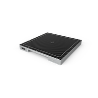
이 카테고리의 Thiết bị hình ảnh điện tử y tế는 전문 의료 영상 및 일부 특수 영상 환경에서 사용되는 플랫 패널 디텍터 중심의 제품군으로 구성되어 있습니다. 특히 DT Detection Technology 제품을 기반으로, Portable DR, Fluoroscopy, O-arm, DSA, Oncology, IGRT 등 서로 다른 운용 목적에 맞는 패널 크기, 센서 기술, 프레임 속도, 인터페이스 구성을 비교할 수 있습니다.
플랫 패널 디텍터가 의료 영상에서 중요한 이유
플랫 패널 디텍터는 X-ray 에너지를 디지털 영상 데이터로 변환하는 핵심 부품입니다. 영상 선명도, 데이터 획득 속도, 시스템 응답성은 물론, 장비 전체의 통합 방식에도 직접적인 영향을 주기 때문에 의료 영상 장비 선정에서 매우 중요한 위치를 차지합니다.
실제 도입 관점에서는 단순히 “해상도가 높은가”만으로 결정하기 어렵습니다. 촬영 목적이 단일 촬영인지, 연속 프레임이 필요한지, 이동형 장비인지 고정형 장비인지에 따라 적합한 디텍터 특성이 달라지므로, 카테고리 전체를 하나의 영상 솔루션 생태계로 보는 접근이 더 실용적입니다.
센서 기술과 변환층은 어떻게 봐야 하나
등록된 제품에는 a-Si-TFT 기반 모델과 IGZO-TFT 기반 모델이 함께 포함되어 있습니다. 일반적으로 이 차이는 영상 획득 속도, 시스템 설계 방향, 적용 분야를 함께 고려해 해석해야 하며, 센서 기술만 따로 떼어 놓고 우열을 판단하는 방식은 적절하지 않을 수 있습니다.
또한 CsI와 DRZ-Plus 같은 scintillator 구성도 확인할 필요가 있습니다. 이 층은 X-ray 신호가 디지털로 읽히기 전에 어떤 방식으로 변환되는지와 관련되며, 실제 영상 특성과 용도 적합성에 영향을 줍니다. 다수의 모델이 140 µm 픽셀 피치를 사용하고, 일부 제품은 100 µm까지 지원해 더 세밀한 영상 표현이 필요한 환경에서 검토할 수 있습니다.
적용 분야별로 보는 제품 선택 포인트
Portable DR와 같이 이동성과 범용 촬영이 중요한 경우에는 패널 면적과 해상도 균형이 중요합니다. 예를 들어 DT Detection Technology X-Panel 4343a PSM-CSV는 430 x 430 mm급 액티브 영역과 3072 x 3072 구성을 제공하며, X-Panel 4343a PSM-CSV-X는 같은 430 x 430 mm급 패널에서 4288 x 4288, 100 µm 구성을 통해 보다 세밀한 영상 확보를 고려하는 프로젝트에 적합한 비교 대상이 됩니다.
반면 연속 촬영이나 고속 영상이 필요한 Fluoroscopy, O-arm, DSA 환경이라면 X-Panel 4343z FPM-CSV-TG 또는 X-Panel 4343z FDM-CSV-TG처럼 10 G Ethernet 기반의 고속 전송 모델을 우선적으로 검토하는 것이 자연스럽습니다. 치료 및 특수 시스템 쪽에서는 X-Panel 4343a FQM-GPG-UH가 Oncology, IGRT 용도와 연결되며, X-Panel 4343a FRM-CSV는 Digital Radiography와 Fluoroscopy 워크플로를 함께 고려해야 하는 시스템에서 참고할 수 있습니다.
패널 크기와 해상도는 함께 비교해야 합니다
이 카테고리에는 430 x 430 mm급 대형 포맷과 350 x 427 mm급 비교적 컴팩트한 포맷이 함께 포함되어 있습니다. 대형 포맷은 넓은 촬영 범위를 요구하는 환경에 유리할 수 있고, 상대적으로 작은 포맷은 장비 외형, 무게, 설치 공간, 기구 설계 제약을 고려해야 하는 프로젝트에서 장점이 있습니다.
예를 들어 X-Panel 3543a PSI-CSV, X-Panel 3543a PSI-GPG, X-Panel 3543a PSI-CSV-T는 350 x 427 mm급 구성을 보여주는 대표적인 예입니다. 반대로 430 x 430 mm급 제품군은 더 넓은 액티브 영역을 바탕으로 시스템 설계 폭을 넓혀줍니다. 해상도 역시 단순한 숫자 비교보다 픽셀 매트릭스와 픽셀 피치를 함께 봐야 하며, 3072 x 3072 / 140 µm와 4288 x 4288 / 100 µm는 각각 지향하는 영상 목적이 다를 수 있습니다.
인터페이스와 프레임 속도는 통합 난이도를 좌우합니다
데이터 인터페이스는 설치 방식과 대역폭, 처리 속도에 직접 연결됩니다. 많은 모델이 Gigabit Ethernet을 사용하고 있어 고정형 또는 이동형 디지털 영상 시스템에서 폭넓게 검토할 수 있으며, 비교적 표준적인 네트워크 기반 통합에 적합합니다.
반면 실시간성 또는 준실시간성이 중요한 경우에는 10 G Ethernet 지원 여부가 더 중요해집니다. X-Panel 4343z FDM-CSV-TG는 높은 프레임 속도를 통해 Fluoroscopy, O-arm, DSA 같은 동적 영상 환경에 적합한 방향성을 보여주며, X-Panel 4343a FQM-GPG-UH는 Optical Fiber 인터페이스를 제공해 전송 환경이나 설치 구조 측면에서 별도의 요구가 있는 시스템에 참고할 수 있습니다.
기계적 사양과 환경 조건도 반드시 확인해야 합니다
실제 산업 및 병원 장비 통합에서는 영상 성능만큼이나 크기, 두께, 무게, 보호 등급이 중요합니다. 예를 들어 일부 3543 시리즈는 상대적으로 가볍고 얇은 설계를 보여 이동형 또는 공간 제약이 있는 설계에 유리할 수 있으며, 반대로 더 무겁고 두꺼운 패널은 고정형 또는 고성능 시스템의 일부일 가능성이 높습니다.
IP 등급, 동작 온도, 습도 조건 역시 장기 운용성 판단에 필요합니다. 대부분의 모델은 IP41 또는 IPX0 수준으로 제시되지만, X-Panel 4343a FQM-GPG-UH는 IP65로 구분되어 더 까다로운 운용 환경을 고려하는 프로젝트에서 눈여겨볼 수 있습니다. 이런 요소는 유지보수 계획과 장비 수명 예측에도 영향을 줍니다.
구매 검토 시 실무적으로 확인할 항목
디텍터를 선택할 때는 먼저 정지 영상용인지 동적 영상용인지, 필요한 액티브 영역이 어느 정도인지, 원하는 세부 표현 수준이 어느 정도인지, 그리고 호스트 시스템이 어떤 인터페이스를 수용할 수 있는지를 정리하는 것이 좋습니다. 그다음 PSI, PSM, PRM, FRM, FPM, FDM, FQM 계열이 각각 어떤 용도에 더 가까운지 비교하면 선택 범위를 훨씬 효율적으로 좁힐 수 있습니다.
의료 장비 도입을 더 넓은 관점에서 검토한다면, 보완적인 계측 장비 카테고리도 함께 살펴보는 것이 유용할 수 있습니다. 예를 들어 임상 장비 구성을 확장하는 경우 혈압 모니터, 혈당 측정기, Medical Thermometer 같은 연관 카테고리도 함께 검토할 수 있습니다.
자주 묻는 질문
정지 영상용 디텍터와 동적 영상용 디텍터의 차이는 무엇인가요?
정지 영상용은 단일 촬영 중심으로 보는 경우가 많고, 동적 영상용은 Fluoroscopy나 DSA처럼 연속 프레임 획득이 필요한 환경에 더 적합합니다. 따라서 프레임 속도와 인터페이스 요구사항이 크게 달라질 수 있습니다.
픽셀 피치가 작을수록 항상 더 좋은가요?
반드시 그렇지는 않습니다. 픽셀 피치가 작으면 더 세밀한 표현에 유리할 수 있지만, 실제 선택은 촬영 목적, 패널 크기, 시스템 비용, 처리 성능까지 함께 고려해야 합니다.
해상도만 보고 선택해도 될까요?
아닙니다. 액티브 영역, 프레임 속도, 인터페이스, 무게, 두께, 보호 등급 같은 조건도 시스템 적합성에 큰 영향을 주므로 종합적으로 비교하는 것이 바람직합니다.
의료 영상용 디텍터를 검토할 때는 숫자가 높은 모델을 고르는 방식보다, 실제 워크플로와 통합 조건에 맞는 구성을 찾는 접근이 더 안정적입니다. 이 카테고리는 다양한 영상 목적에 대응할 수 있는 플랫 패널 디텍터를 중심으로 구성되어 있으므로, 적용 분야와 시스템 요구사항을 먼저 정리한 뒤 제품군을 비교하면 보다 실질적인 선택에 도움이 됩니다.
인증된 전문가에게 직접 연결